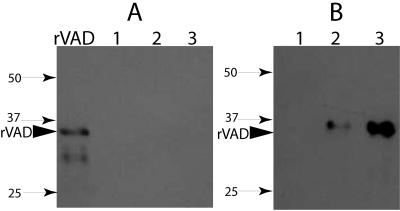
FIG. 2.
Detection of SpoVAD in the coat-outer membrane fraction (A) and disrupted decoated spores of B. subtilis strains (B). Samples of the coat-outer membrane extract (A) or the total extract from disrupted decoated spores of various strains (B) were run on SDS-PAGE and analyzed by Western blotting as described in Materials and Methods. The samples run in the various lanes were from 70 μg dry spores of strains PS3406 (sleB spoVA) (lane 1), PS832 (wild-type) (lane 2), and PS3411 (PsspB::spoVA) (lane 3). The arrowhead denotes the migration position of the SpoVAD fusion protein, and the numbered arrows denote the migration positions of molecular mass markers (sizes are in kilodaltons).

