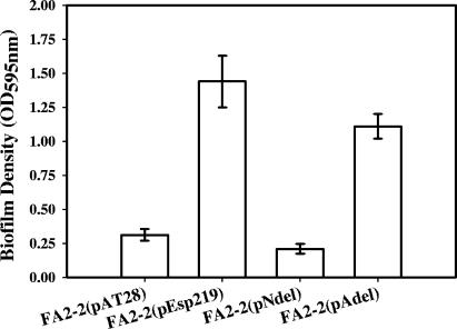
FIG. 2.
Biofilm formation by Esp-negative FA2-2(pAT28), Esp-positive FA2-2(pEsp219), Esp N-terminal deletion mutant FA2-2(pNdel), and Esp A-repeat deletion mutant FA2-2(pAdel) grown in TSB supplemented with 0.75% glucose. Biofilm formation was quantified by crystal violet staining followed by extraction of bound crystal violet into an ethanol-acetone mixture. The y axis represents the optical densities of dissolved crystal violet measured at 595 nm. The error bars represent means ± standard errors.

