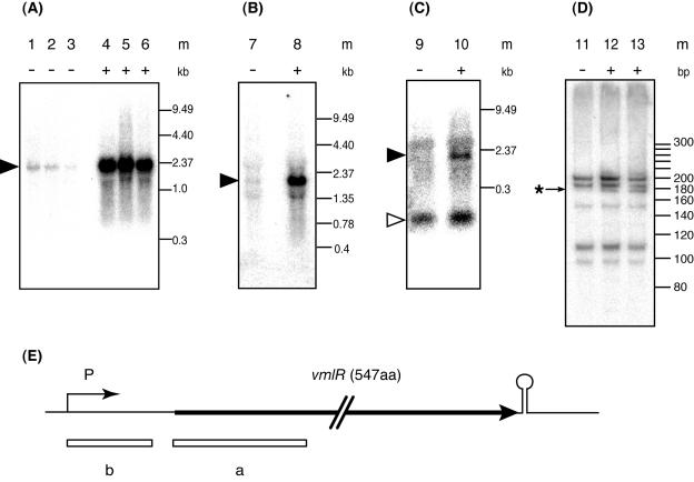
FIG. 2.
Induction of vmlR expression by the addition of virginiamycin M or lincomycin in growth medium. An overnight culture of B. subtilis 168 grown in LB medium was inoculated into LB medium (optical density at 530 nm of 0.05) without or with virginiamycin M or lincomycin. Samples were withdrawn during early (optical density at 530 nm of 0.5; lanes 1, 4, 7, 8, 9, 10, 11, 12, and 13), middle (optical density at 530 nm of 1.2; lanes 2 and 5) and late (optical density at 530 nm of 2.0; lanes 3 and 6) log-phase growth. Culture media were the following: LB (lanes 1, 2, 3, 7, 9, and 11), LB containing 1.9 μg ml−1 virginiamycin M (lanes 4, 5, 6, 10, and 12), and LB containing 2 μg ml−1 lincomycin (lanes 8 and 13). m, molecular size standard. Total RNA (5 μg) was loaded per lane. For Northern blotting, a 1.5% agarose gel containing 6% formaldehyde (A, B, and C) and a 6% polyacrylamide gel containing 7 M urea (D) were used. Probe A containing sequences from +213 to 493 was used in panels A and B; probe B containing sequences from +3 to 181 (+1, transcription initiation site) was used in panels C and D. A filled arrow indicates the 1.9-kb vmlR transcript, and an open arrow indicates transcripts less than 300 bp in length. An asterisk indicates an extra band detected under the induced condition. (E) Thick arrow, open reading frame of vmlR; P, promoter; circle on stem, transcriptional terminator; boxes a and b under the open reading frame, probes.

