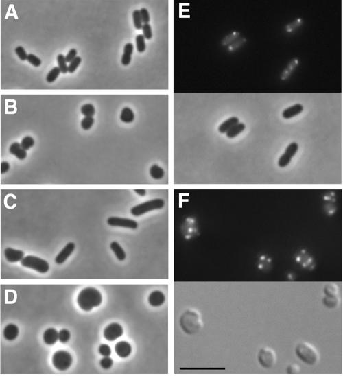
FIG. 2.
Requirement of mreBCD for partial complementation of morphological and IcsA localization phenotypes of mreB cells. Bright-field (panels A to D and E, bottom), DIC (panel F, bottom), and fluorescence (panels E and F, top) microscopy of live E. coli mreB cells, with or without complementation with mreB or mreBCD in trans. (A) Rod-shaped wild-type strain. (B) Spherically shaped MC1000 ΔmreB::FRT-cat-FRT. (C) MC1000 ΔmreB::FRT-cat-FRT expressing mreBCD in trans. (D) MC1000 ΔmreB::FRT-cat-FRT expressing mreB in trans. (E) MC1000 ΔmreB::FRT-cat-FRT expressing mreBCD and IcsA507-620-GFP. (F) MC1000 incubated with the MreB inhibitor A22 for 2 h and expressing IcsA507-620-GFP. Size bar, 5 μm.

