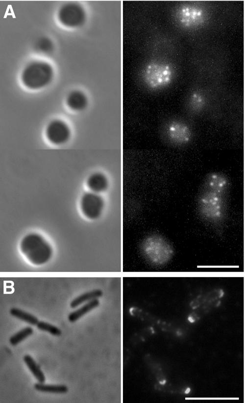
FIG. 4.
Distribution of full-length secreted IcsA on the surface of mreB cells. Shown are results from immunofluorescence (right panels) and bright-field (left panels) microscopy of intact cells expressing full-length secreted IcsA. Derivatives of E. coli strain 2443, which contains an intact LPS, are depicted. Detection of surface IcsA was by indirect immunofluorescence using antibody to the extracellular domain of IcsA. (A) 2443 mreB::Tn10 ompT expressing IcsA. Because the signal was weak, the images were adjusted to enhance the intensity of the fluorescent signal and its contrast with the background signal. (B) 2443 ompT expressing IcsA. Size bars, 5 μm.

