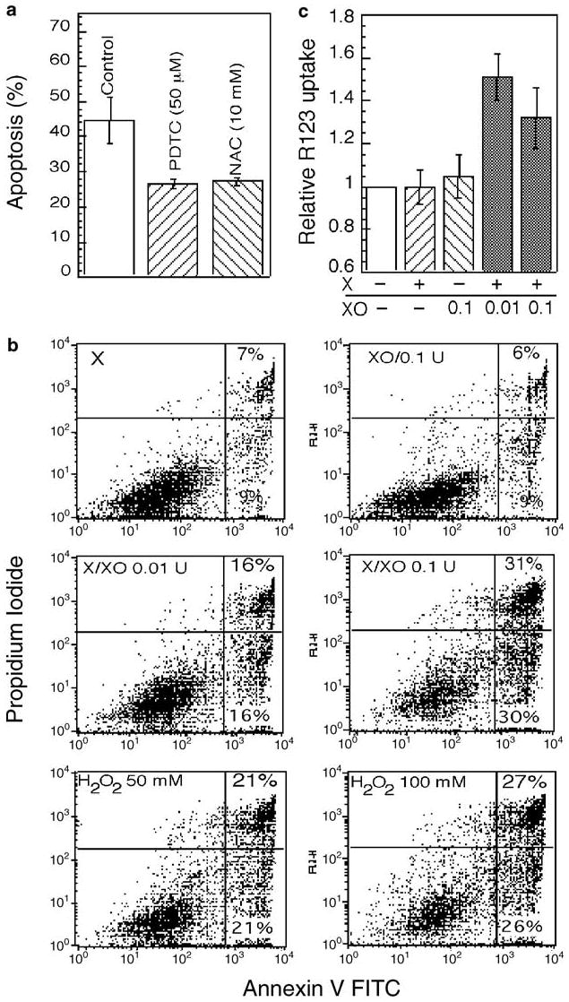
Figure 5.

Effect of anti- and pro-oxidants on apoptosis and Δψm. (a) Morphological changes characteristic of apoptosis were determined by DAPI staining in cells pretreated with the antioxidants PDTC (50 μM) and NAC (10 mM) for 1 h before irradiaton (10 Gy). Apoptosis was then examined at 28 h after IR. The data represent mean values from three separate experiments. (b, c) The effect of xanthine (X) and xanthine oxidase (XO) on apoptosis and Δψm was examined in cells treated with X and XO, alone or in combination, or H2O2 for 30 min at 30°C and then subjected to R123 staining (c) or incubated in complete medium for 24 h before annexin V staining and flow cytometry (b)
