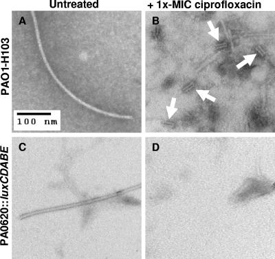
FIG. 2.
Electron micrographs of supernatants from P. aeruginosa cells that were untreated or treated with 1× MIC of ciprofloxacin. (A) Untreated strain H103 cells. (B) 1× MIC of ciprofloxacin-treated strain H103 cells. Arrows point to the presence of pyocin/phage tail structures. (C) Untreated strain PA0620::luxCDABE cells. (D) 1× MIC of ciprofloxacin-treated PA0620::luxCDABE cells showing absence of R-type pyocin tail structures.

