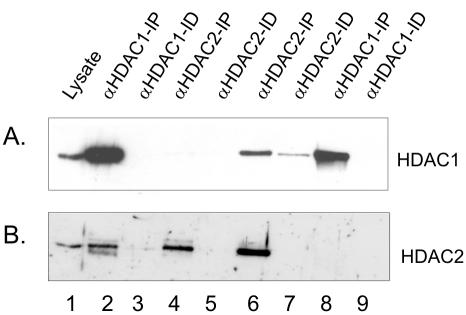
Figure 2.
Coimmunoprecipitation of HDAC1 and HDAC2. Two A260 of MCF-7 cell lysate was incubated with 4 μg of anti-HDAC1 antibodies, and the immunoprecipitated (IP, lane 2) and immunodepleted (ID, lane 3) fractions were collected. The immunodepleted fraction was next incubated with anti-HDAC2 antibodies, yielding IP (lane 4) and ID (lane 5) fractions. Conversely, two A260 of MCF-7 cell lysate were incubated with 4 μg of anti-HDAC2 antibodies, and the immunoprecipitated (IP, lane 6) and immunodepleted (ID, lane 7) fractions were collected. The immunodepleted fraction was next incubated with anti-HDAC1 antibodies, yielding IP (lane 8) and ID (lane 9) fractions. The whole IP fractions and equivalent volumes of lysate (lane 1) and ID fractions, corresponding to 1% of 2 A260 of lysate were loaded onto SDS-10% polyacrylamide gels, transferred to nitrocellulose membranes, and immunochemically stained with anti-HDAC1 (A) or anti-HDAC2 (B) antibodies.

