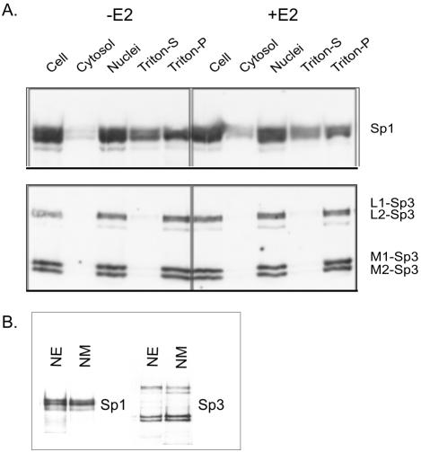
Figure 8.
Subcellular distribution of Sp1 and Sp3 in MCF-7 cells. (A) MCF-7 cells incubated with (+E2) or without (–E2) 10 nM estradiol at 37°C for 20 min were fractionated by Triton X-100 extraction. Equal volumes of all fractions were analyzed by SDS- 10% PAGE, transferred to a membrane, and immunochemically stained with anti-Sp1 (top) or anti-Sp3 antibodies (bottom). The long isoforms of Sp3 are shown as L1 and L2, whereas the short isoforms are denoted by M1 and M2. (B) Association of Sp1 and Sp3 with nuclear matrix: 20 μg of protein of nuclear extract (NE) and nuclear matrix (NM) from MCF-7 cells, grown in estrogen complete medium, were electrophoresed on a SDS-10% polyacrylamide gel, and immunochemically stained with anti-Sp1 (left) or anti-Sp3 (right) antibodies.

