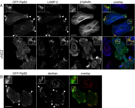
Figure 5.
Functionality of the redistributed LE/Lys. (A) Positioning of GFP-PipB2 and redistributed LE/Lys requires an intact microtubule network. HeLa cells were transiently transfected with pGFP-PipB2 for 24 h and either left untreated (top) or treated with 5 μg/ml NDZ for 30 min (+NDZ, bottom). Monolayers were fixed, permeabilized, and immunostained for β-tubulin and LAMP-2 and viewed by confocal microscopy. An overlay of the three channels is presented on the right (GFP-PipB2, green; β-tubulin, blue; and LAMP-2, red). Insets show 2× enlargement of boxed area. Arrowheads indicate peripheral accumulation of GFP-PipB2 and LAMP-2. Bar, 20 μm. (B) The redistributed LE/Lys are accessible to fluid phase markers. HeLa cells were transiently transfected with pGFP-PipB2 and then incubated for 12 h with 130 μg/ml Alexa Fluor 568-labeled dextran, washed, and further incubated in dextran-free medium for 1 h to label LE/Lys. Monolayers were fixed and examined by confocal microscopy. Arrowheads indicate peripheral accumulation of dextran and GFP-PipB2 in transfected cells. An overlay of the two channels is shown on the right (GFP-PipB2, green; and dextran, red). Insets show 2× enlargement of boxed area. Bar, 20 μm.

