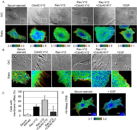
Figure 6.
Effect of Cdc42 on the distribution of RhoA activity. (A) HeLa cells expressing Raichu-RhoA with or without expression vectors, indicated at the top of each panel, were imaged for YFP, CFP, and DIC. Representative pseudocolor images of the Ratio (FRET efficiency) and DIC images are shown. The rightmost panels show EGF-stimulated cells as a control. (B) Magnified images of the peripheral regions boxed in white in A. (C) The appearance of membrane ruffling was scored. The values are presented as percentages of total transfected cells. The data were averaged over three independent experiments and are shown with the SD (D) Cos1 cells expressing Raichu-RhoA and CRIB domain of N-Wasp was stimulated with EGF as in Figure 2.

