Figure 8.
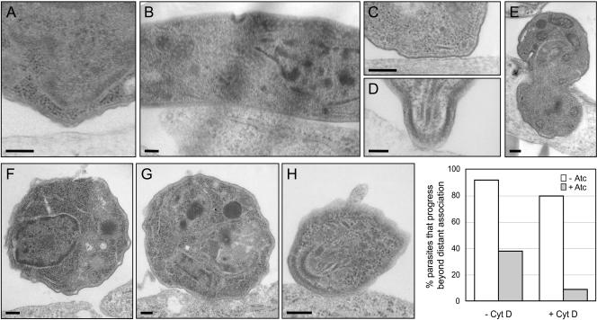
Ultrastructural characterization of the interaction between TgAMA1-depleted parasites and host cells. Δama1/AMA1-myc parasites were incubated with host cells for 1 min at 37°C, fixed, and examined by transmission electron microscopy. Representative images of parasites, grown with (A) or without (B-E) Atc and in various states of interaction with host cells, are shown. In most profiles of TgAMA1-depleted tachyzoites examined, the parasite had not progressed beyond the distant attachment step (A). In contrast, the same parasites grown in the absence of Atc were frequently observed in intimate lateral (B) or apical (C and D) association with the host cell membrane or were found partially (E) or fully (unpublished data) internalized. When the parasites were pretreated with CytD before addition to the host cells, the majority of the Atc-treated, TgAMA1-depleted parasites again had not progressed beyond the initial attachment phase in the profiles examined (e.g., F), whereas most of those grown in the absence of Atc progressed to the point of forming intimate (G), often apical (H), associations with the host cell. Scale bars, 200 nm. The percentage of Δama1/AMA1-myc parasites (+/- Atc) that progress beyond distant association with the host cell, with or without CytD pretreatment, is shown in the graph.

