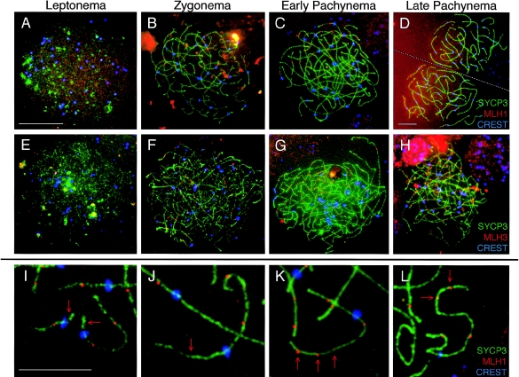
Figure 5.
Localization of MLH1 and MLH3 during prophase I in human fetal oocytes. The MutL homologs, MLH1 and MLH3, localize to human female meiotic chromosomes during each substage of prophase I. A–D, MLH1 (red, TRITC) is shown with SYCP3 (green, FITC) and CREST (blue, Cy5) during leptonema (A), zygonema (B), early pachynema (C), and late pachynema (D). E–H, During each stage, MLH3 (red, TRITC) is shown with SYCP3 (green, FITC) and CREST (blue, Cy5). E, Leptonema; F, zygonema; G, early pachynema; and H, late pachynema. I–L, High magnification images of pachytene chromosomes highlights extreme variability in MLH1 localization, with some chromosome arms of pachytene oocytes often having too few MLH1 foci (arrows in I and J) or too many MLH1 foci (arrows in K and L). Green, SYCP3; red, MLH1; blue, CREST. Scale bar for panels A–C and E–H = 10 μm, scale bar for panel D = 10 μm, and scale bar for panels I–K = 5 μm. Note: at leptonema, γH2AX localization is almost continuous along the developing SC, so counts at this stage do not necessarily represent single foci.

