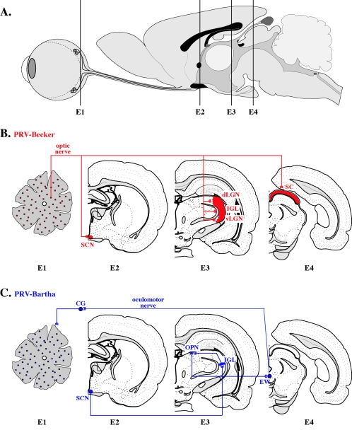
FIG. 5.
Rat eye infection model: comparison of wild-type (PRV-Becker) and attenuated (PRV-Bartha) neuronal spreads. A. Sagittal view of the rat brain. E1, E2, E3, and E4 refer to the level of coronal sections depicted in panels B and C. B. Spread of PRV-Becker (shown in red) after virus injection into the vitreous humor of the rat eye. PRV-Becker spreads from infected retinal ganglion cells (level E1) through the optic nerve to second-order neurons in the supracharismatic nucleus (level E2), dorsal and ventral aspects of the geniculate nuclei (dorsal aspect of the lateral geniculate nucleus and ventral aspect of the lateral geniculate nucleus) and intergeniculate nucleus (level E3), and the superior colliculus of thevisual cortex (level E4). Only first-order projections are shown. C. Transport of PRV-Bartha (shown in blue) is restricted to retrograde-only pathways. Although cells of the retinal ganglia (level E1) are infected with PRV-Bartha, this virus is restricted from anterograde spread through the optic nerve to retinorecipient neurons. Instead, retrograde spread of infection to first-order neurons in the ciliary ganglion leads to transport through the oculomotor nerve to second-order neurons in the Edinger-Westphal nucleus (level E4). Infection of neurons in the olivary pretectal nucleus, intergeniculate nucleus (level E3) and supracharismatic nucleus (level E2) is also by retrograde axonal transport of virus as shown (369). Abbreviations: CG, ciliary ganglion; dLGN, dorsal aspect of the lateral geniculate nucleus; EW, Edinger-Westphal nucleus; IGL, intergeniculate nucleus; OPN, olivary pretectal nucleus; SC, superior colliculus; SCN, suprachiasmatic nucleus; vLGN, ventral aspect of the lateral geniculate nucleus. (Figure modified from reference 386 with permission of the publisher.)

