
Figure 5.
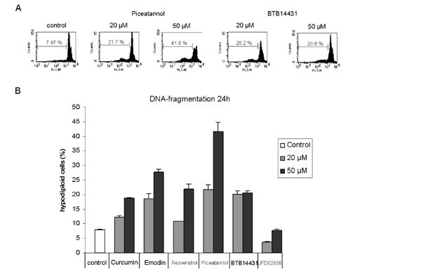
Detection of DNA-fragmentation in HeLa cells after 24 h incubation with kinase inhibitors. Dose-dependent DNA-fragmentation in HeLa cells after 24 h treatment with curcumin- and emodin-derived inhibitors. (A) Representative data for piceatannol (20 μM and 50 μM) and BTB14431 (20 μM and 50 μM) are shown. As a control the cells were incubated with DMSO (0.1%). Piceatannol and BTB14431 were added in two concentrations (20 μM and 50 μM). The marker indicates the percentage of subG1 cells (apoptotic cells). (B) The bar chart shows the percentage of hypodiploid cells (apoptotic cells) after treatment with the inhibitors curcumin, emodin, resveratrol, piceatannol, BTB14431 or JFD02836 at different concentrations (20 μM and 50 μM).