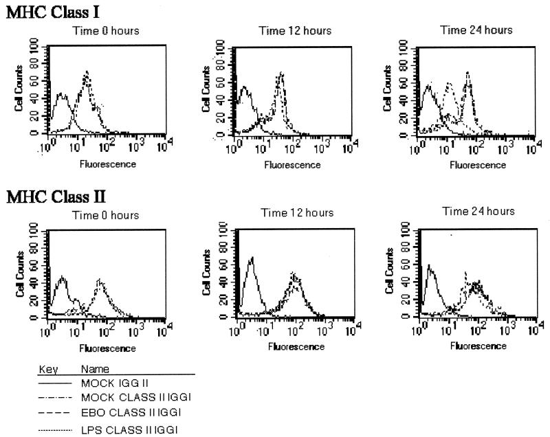
FIG. 2.
HLA-A, B, C, and HLA-DR expression by alveolar macrophages. Alveolar macrophages from cynomolgus monkeys exposed to medium (mock-infected control), Ebola virus (EBO) (MOI, 1.0), or LPS were harvested at 0, 12, and 24 h postinfection. Cells were left untreated or were treated with monoclonal antibodies to HLA-DR (MHC class II) or to HLA-A, B, and C (MHC class I), washed, fixed in 4% paraformaldehyde, and analyzed by fluorescence-activated cell sorting. A total of 104 cells per condition were analyzed. Relative fluorescence intensity for HLA-DR and for HLA-A, HLA-B, and HLA-C proteins is displayed on the horizontal axis. These data are from a representative experiment with cells from one of the four cynomolgus monkeys.

