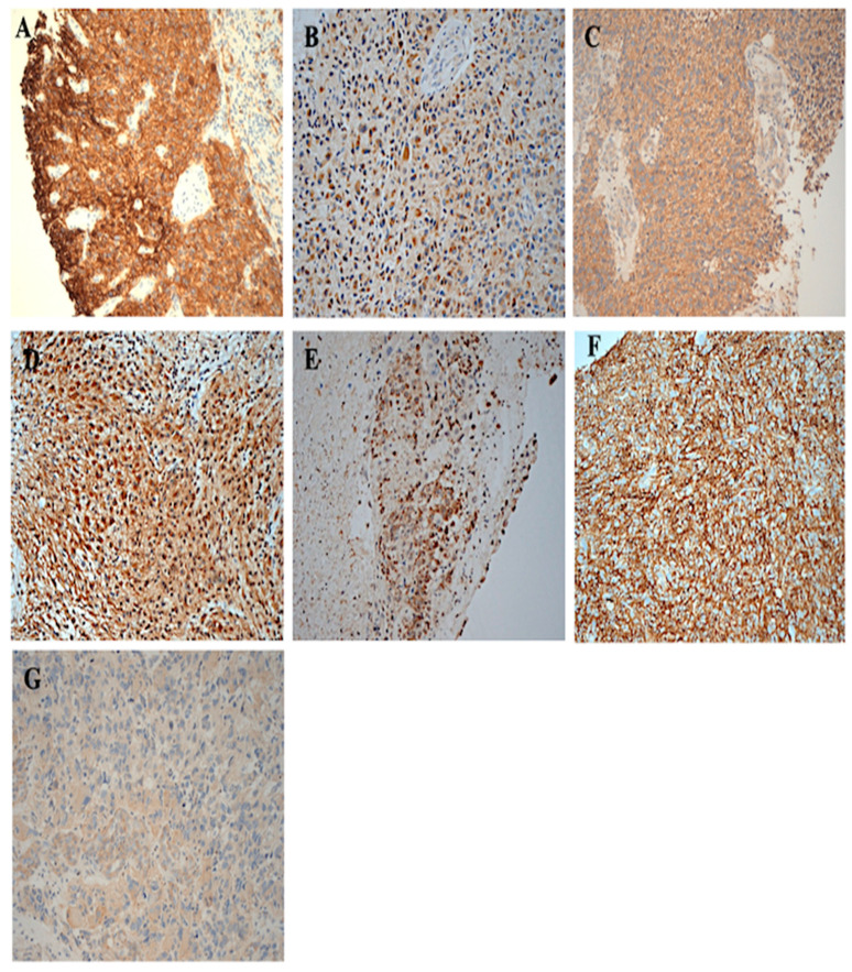
Figure 1.
The immunohistochemical staining of whole brain tumour specimens from patients with glioblastoma for the expression of the HER-family members. Formalin-fixed paraffin-embedded tumour sections were stained with primary mAb as described under Materials and Methods. (A) EGFR 3+, cytoplasmic/membranous, ×200 magnification (B) HER2 2+ cytoplasmic, ×200 magnification (C) HER3 2+ cytoplasmic, ×200 magnification (D) HER4 2+ cytoplasmic, ×200 magnification (E) EGFRvIII 2+ cytoplasmic, (F) CD44 3+ membranous, (G) CD109 1+ cytoplasmic, ×200 magnification.

