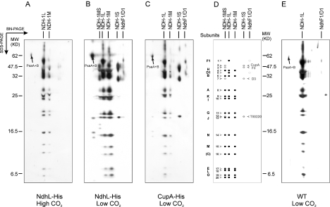
Figure 3. Two-dimensional gels of NDH-1 complexes purified on the Ni2+ affinity column.
Thylakoid membranes were isolated, solubilized with 0.25% β-DM, and applied to the Ni2+ affinity column. Eluted complexes were subjected to BN/SDS/PAGE, and the gels were stained with silver (A, B, C and E): (A) NdhL-His strain grown at high CO2; (B) NdhL-His strain grown at low CO2; (C) CupA-His strain grown at low CO2; (D) scheme of NDH-1 subunits; (E) WT grown at low CO2. The identify of the spots is based on MS analysis and as indicated in Tables 1 and 2.

