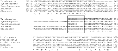
Figure 4. Sequence comparison of the NdhF1 proteins from cyanobacteria.
Multiple sequence alignment of the NdhF1 protein on the region surrounding the proteolytic site and the probable Ni2+-affinity-column-binding site was performed by ClustalW. Sequences of the following organisms were used: T. elongatus (T. elongatus BP-1), Tll0720; Synechocystis (Synechocystis sp. PCC 6803), Slr0844; Prochlorococcus (Prochlorococcus marinus SS120), Pro0172; Anabaena (Anabaena sp. PCC 7120), Alr3956; Gleobacter (Gleobacter violaceus PCC 7421), Glr0218. The arrow indicates the proteolytic site in Synechocystis 6803 NdhF1 protein. The box indicates the probable Ni2+-affinity-column-binding site, and the grey shading shows the histidine-rich region in T. elongatus.

