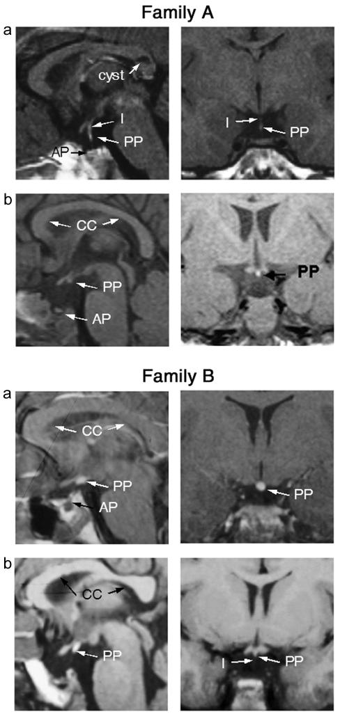
Figure 1.
Family A, MRI scans in patients with SOX3 duplication. Coronal and sagittal MRI scans of patient 1 (panel a) and patient 2 (panel b) from family A, showing APH (“AP”), partial hypoplasia of the infundibulum (“I”) in patient 1 (absent in patient 2), and an undescended/ectopic posterior pituitary (“PP”) (partial in patient 1). Note the cyst in the corpus callosum identified in patient 1. Family B, MRI scans in patients with PA expansion in SOX3. Coronal and sagittal MRI scans of patient 2 (panel a) and patient 3 (panel b) from family B, showing APH (“AP”), hypoplasia of the infundibulum (“I”), and an undescended/ectopic posterior pituitary (“PP”).

