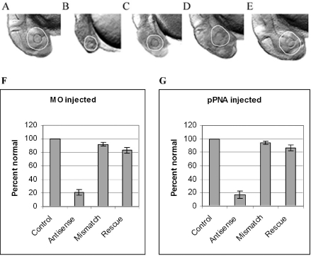
Figure 7.
Sense mRNA rescue of ccnd1 antisense-treated zebrafish embryo morphology. Zebrafish embryos were microinjected with 0.5 mM ccnd1 antisense MO or HypNA-pPNA as described in Materials and Methods. Sense ccnd1 mRNA at 1 g/l was co-microinjected with the either MO or HypNA-pPNA into selected groups of embryos (designated ‘rescue’), and morphology recorded at 24 h after injection: (A) Phenol red-treated control; (B) MO-treated embryo; (C) HypNA-pPNA-treated embryo; (D) MO plus mRNA co-injected embryo; (E) HypNA-pPNA plus mRNA co-injected embryo; all images were taken at ×100 magnification. All embryos were treated with PTU following microinjection to inhibit pigment formation; therefore, the circumference of the eyes are delineated for better visualization. There is visual evidence of rescue of the ccnd1 antisense-induced microophthalmia and microcephaly in the ccnd1 mRNA co-injected embryos. The incidence of microophthalmia in MO- (F) or HypNA-pPNA-treated (G) embryos, with or without sense mRNA rescue, was calculated and expressed as percent (±SD) of 100 embryos per sample.

