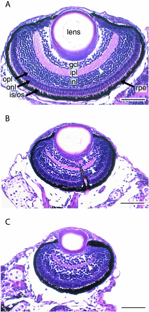
Figure 8.
Histologic appearance of zebrafish eyes following ccnd1 knockdown. Representative coronal sections of the retinas of 5-day-old larvae are shown. (A) Phenol red-treated control. (B) HypNA-pPNA-treated embryo. (C) MO-treated embryo. Layers are indicated as follows: GCL, ganglion cell layer; INL and ONL, inner and outer nuclear layer, respectively; IPL and OPL, inner and outer plexiform layer, respectively; RPE, retinal pigmented epithelium. White arrow heads indicate thickness of the inner plexiform layer. Scale bar: 50 µm.

