Figure 4.
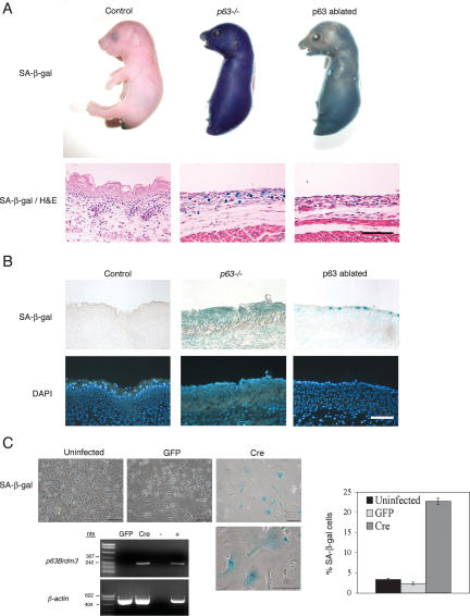
The senescence marker SA-β-gal is induced in response to both germline and somatically induced p63 deficiency in vivo, and in primary keratinocytes in culture. (A) A whole-mount assay for endogenous SA-β-gal activity was performed on E17.5 p63+/+, p63-/-, and p63-ablated embryos; skin sections from SA-β-gal assayed embryos were subjected to histological analysis; the intense blue color observed in p63-/- embryos is indicative of senescent cells. Bar, 100 μM. (B) Dorsal back skin biopsies from E17.5 control, p63-/-, and p63-ablated embryos (treated with mifepristone at E8.5) were stained with Dapi to show histology (bottom panels) and assayed for endogenous SA-β-gal activity (top panels). Positive (blue) cells were essentially undetectable in control skin, whereas p63-/- and p63-ablated skin had a dramatic increase in cellular senescence. Bar, 100 μM. (C) Primary keratinocyte cultures from p63flox/flox mice were uninfected, or infected with GFP-expressing (GFP) or Cre-expressing (Cre) retrovirus. DNA prepared from these cultures was subjected to PCR to identify the disrupted p63Brdm3 allele. β-Actin was used as a positive control. Cultures were subjected to the SA-β-gal assay, and senescent cells were quantitated. Bar, 100 μm.

