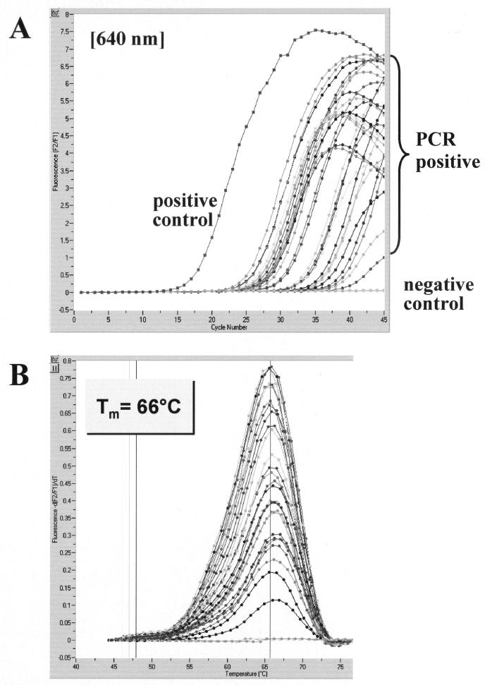
FIG. 2.
Evaluation of the LightCycler PCR assay with brain tissue sections of C. neoformans-infected mice. A representative set of 28 samples with various C. neoformans concentrations was tested for the presence of C. neoformans 18S rDNA. Amplicon curves representing C. neoformans PCR-positive isolates are indicated by braces (A), and the corresponding results in melting curve analysis are depicted (B).

