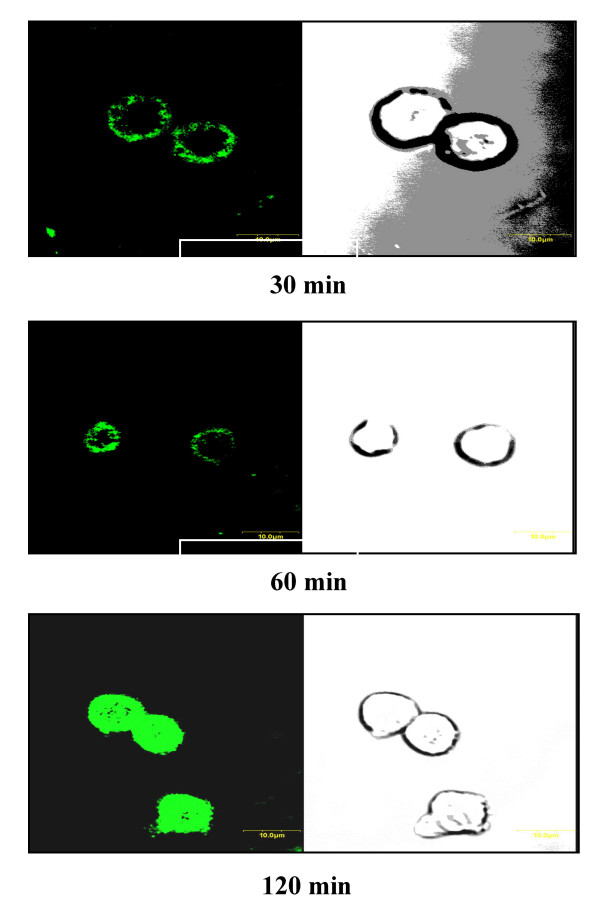
Figure 2.
Confocal microscopy of Jurkat cells after treatment with FITC-labeled Pep27anal2 as a function of time. After incubating FITC-labeled Pep27anal2 for 30, 60, and 120 min, cells were fixed with 3.7% formaldehyde and analyzed using a laser scanning confocal microscope. Pep27anal2 peptide expression is shown as green on the left confocal microphotograph. Cells morphology is shown by the phase contrast microphotograph on the right.

