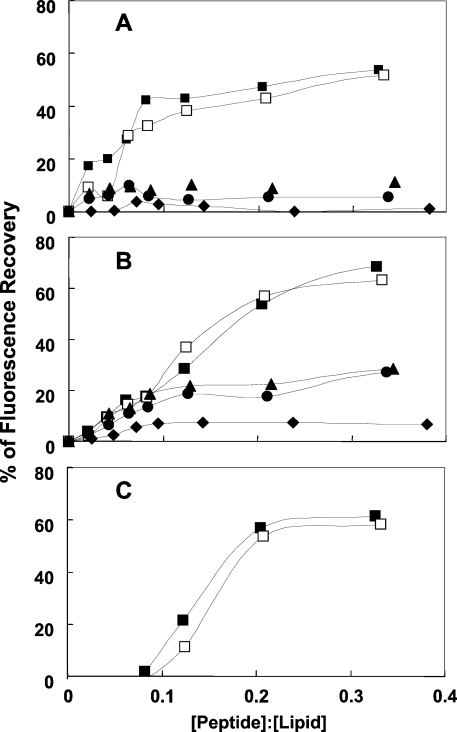
Figure 2. Dose-dependent dissipation of the diffusion potential in vesicles, induced by the lipopeptides.
The lipopeptides were added to isotonic K+-free buffer containing LUVs pre-equilibrated with the fluorescent dyes diS-C3-5 and valinomycin. Fluorescence recovery was measured for 60 min after the peptides were mixed with the vesicles. Lipid concentration was 30 μM and peptide concentration was in the range 0.3–12 μM. (A) PC/cholesterol vesicles, (B) PE/PG vesicles and (C) PC/PE/PI/ergosterol vesicles. Peptide designations: ◆, [D]-L6K6; ▲, [D]-L6K6DA; ●, [D]-L6K6-DDA; □, [D]-L6K6-MA; and ■, [D]-L6K6-PA.

