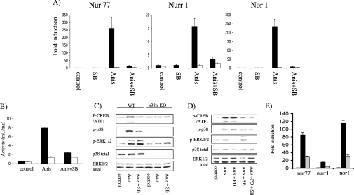
Figure 4. ERK1/2 and p38α are required for NR4A induction by anisomycin.
Immortalized p38α knockout (white bars) and wild-type (black bars) MEF cells (A–C) were serum-starved for 16 h, incubated with 5 μM SB 203580 (SB) for 1 h where indicated and then left unstimulated (control) or stimulated with 10 μg/ml anisomycin (Anis) for 1 h. After stimulation, total RNA was isolated and the expression of Nur77, Nurr1 and Nor1 determined by real-time PCR (A). Levels of MSK1 activity were determined by immunoprecipitation kinase assay as under the Materials and methods section (B). To determine if the ERK1/2 and p38 pathways were activated, lysates were run on SDS/polyacrylamide gels. The levels of phospho-CREB, phospho-p38, (p-p38), phospho-ERK1/2, total p38α and total ERK1/2 were determined by immunoblotting with specific antibodies (C). (D) Primary wild-type MEF cells were serum-starved for 16 h, incubated for 1 h with 5 μM SB 203580 (SB) and/or 2 μM PD 184352 (PD) where indicated and then stimulated with 10 μg/ml anisomycin (Anis) for 1 h. Cells were then lysed and the levels of phospho-CREB, phospho-p38, phospho-ERK1/2, total p38α and total ERK1/2 were determined by immunoblotting with specific antibodies. (E) Primary wild-type MEF cells were serum-starved for 16 h, incubated for 1 h with 10 μM U0126 (grey bars) or DMSO control (black bars) and then stimulated with 10 μg/ml anisomycin for 1 h. The induction of Nur77, Nurr1 and Nor1 mRNA was then determined by real-time PCR as described in the Materials and methods section. Error bars represent the S.E.M. of three individual stimulations.

