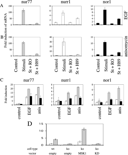
Figure 6. Expression of MSK1 in MSK1/MSK2−/− cells rescues NR4A expression.
(A) Wild-type immortalized MEF cells were serum-starved for 16 h and then incubated with 5 μM Ro 318220 (RO) or 10 μM H89 were indicated. Cells were then left unstimualed, or stimulated (St) with EGF (100 ng/ml) for 1 h. The induction of Nur77, Nurr1 and Nor1 mRNA was measured by real-time PCR as described in the Materials and methods section. Error bars represent the S.E.M. of three individual stimulations. (B) As in (A) except that cells were stimulated with anisomycin (10 μg/ml) for 1 h. (C) Immortalized MSK1/MSK2−/− MEF cells were transfected with either empty vector (white bars) or wild-type (light grey bars) or kinase-dead (black bars) MSK1 expression vectors. Cells were then serum-starved for 16 h and left unstimulated (control), or stimulated with either EGF (100 ng/ml) or anisomycin (10 μg/ml), for 1 h. The induction of Nur77, Nurr1 and Nor1 mRNA was measured by real-time PCR as described in the Materials and methods section. Error bars represent the S.E.M. three individual stimulations. (D) Immortalized wild-type (wt) or MSK1/MSK2−/− (ko) MEF cells were transfected with a bp −274 to bp +66 Nur77 promoter luciferase reporter vector, together with either an empty vector, wild-type or kinase-dead (KD) MSK1 expression vector, as indicated. Cells were then serum-starved for 16 h and left unstimulated (white bars), or stimulated with PMA (400 ng/ml) for 1 h. Luciferase activity was measured as described in the Materials and methods section. Error bars represent the S.E.M. of three individual stimulations.

