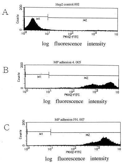
FIG. 2.
Adherence of M pneumoniae No. 4 and FH strains to HEp-2 cells evaluated by flow cytometric analysis. (A) Control HEp-2 cells. (B) HEp-2 cells adhered with PKH-2-labeled M. pneumoniae No. 4 strain. (C) HEp-2 cells adhered with PKH-2-labeled M. pneumoniae FH strain. Adherence of M. pneumoniae was evaluated according to its mean fluorescence intensity.

