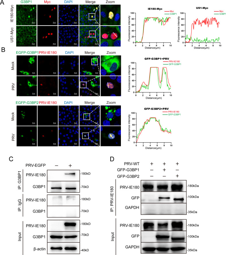
Fig 4.
PRV IE180 interacts with G3BP1/2. (A) IE180-Myc colocalizes with endogenous G3BP1 in the nucleus. PK-15 cells were transfected with IE180-Myc, or empty vector (EV-Myc) and US1-Myc as negative control. After 24 h post-transfection, the cells were fixed and stained with anti-G3BP1 and anti-Myc-tag antibodies, and the nuclei were counterstained with DAPI. Images were taken by a confocal microscope after immunofluorescence. (B) PRV IE180 co-located with GFP-G3BP1/2 in nucleus. PK-15 cells were infected with PRV-WT (MOI = 1) for 12 h after being transfected with GFP-G3BP1 or GFP-G3BP2 for 24 h. The cells were stained with anti-PRV-IE180 antibody, and the nuclei were stained with DAPI. Images were taken by a confocal microscope. (C) PRV IE180 interacts with G3BPs in infected PK-15 cells. PK-15 cells were infected with PRV-EGFP (MOI = 1) for 12 h. Cell lysates were immunoprecipitated with anti-G3BP1 antibody and immunoblotted with either anti-IE180 or anti-G3BP1 antibody. (D) PRV IE180 interacts with GFP-G3BP1/2 in PK-15 cells. PK-15 cells transfected with GFP-G3BP1 or GFP-G3BP2 for 24 h, then infected with PRV-WT (MOI = 1) for 12 h, cell lysates were immunoprecipitated with anti-G3BP1 antibody and immunoblotted with either anti-IE180 or anti-GFP-tag antibodies. The fluorescence colocalization quantification plots were analyzed using ImageJ software. Scale bars = 20 µm.

