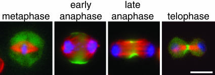
Fig. 1.
Myosin II regulatory light chain-GFP (RLC-GFP) reveals the localization of myosin II during cell division. S2 cells expressing RLC-GFP (green) were fixed and stained for DNA (blue) and α-tubulin (red). RLC-GFP was found in the cytoplasm in metaphase cells but was localized to the midzone cortex at anaphase and through telophase/cytokinesis. (Scale bar: 5 μm.).

