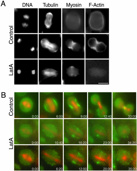
Fig. 2.
F-actin is not required for recruitment of myosin II to the cleavage furrow. (A) S2 cells stably expressing RLC-GFP were treated with DMSO only (Top and Middle) or 20 μM LatA to depolymerize F-actin (Bottom) and then fixed and stained for F-actin, DNA, and α-tubulin. Control cells in early anaphase (Top) show that RLC-GFP is enriched at the midzone cortex earlier than F-actin. Cells treated with LatA (Bottom) did not show F-actin staining, but myosin II is still enriched at the equator. (B) Time-lapse epifluorescence microscopy was used to follow S2 cells stably expressing both RLC-GFP (green) and mRFP-α-tubulin (red) as they transitioned into anaphase. In control cells (Top) RLC-GFP was localized normally at the midzone equator through the final stages of cytokinesis. In cells treated with 20 μM LatA (Middle and Bottom) myosin II is still recruited to the midzone, but it disperses into patches in the membrane after many minutes. (Scale bar: 5 μm.).

