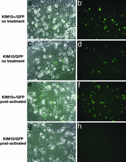
Fig. 1.
Δpgm Y. pestis does not survive in postactivated macrophages. BMM were infected with KIM10+/GFP (pgm+)(a, b, e, and f) or KIM10/GFP (Δpgm) (c, d, g, and h) for 20 min. Gentamicin was then added to prevent survival of extracellular bacteria. The BMM were then left untreated (a- d) or exposed to IFN-γ (e- h; postactivated). After 24 h of infection, isopropyl β-d-thiogalactoside was added to the infected cells to induce GFP expression. One hour later, the samples were fixed and examined by fluorescence or phase microscopy. Representative images were captured by digital photography. Overlays of GFP expression and phase-contrast images are shown (a, c, e, and g).

