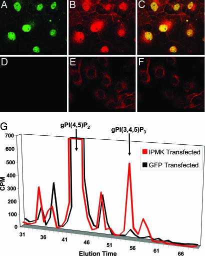
Fig. 4.
In vivo production of nuclear PI(3,4,5)P3 by IPMK. Cos-7 cells transfected with HA-IPMK (A–C) or empty vector (D–F) were fixed and stained via immunofluorescence with anti-HA (A and D, green) and anti-PI(3,4,5)P3 (B and E, red) antibodies. Merged images of cells transfected with HA-IPMK (C) or empty vector (F) reveal an increase in nuclear PI(3,4,5)P3 staining in HA-IPMK-transfected cells that colocalize with nuclear HA-IPMK (overlap shown in yellow). Images were acquired by confocal microscopy. Biochemical identification of increases in PI(3,4,5)P3 in HA-IPMK-transfected cells was accomplished by HPLC analysis (G) of glycerophosphoinositides prepared from [3H]inositol-labeled cells overexpressing HA-IPMK or GFP.

