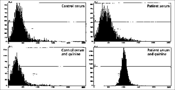
Figure.
Demonstration of quinine-dependent antiplatelet antibodies. The 4 panels demonstrate patterns of flow cytometry analysis, with the number of platelets recorded on the vertical axis and the intensity of brightness of the fluorescence on the horizontal axis. Normal platelets were incubated with normal serum or patient serum, with or without quinine. After the incubation, fluorescein-labeled antihuman immunoglobulin G antibodies were added and flow cytometry performed. In the presence of quinine, antiplatelet antibodies from the patient's serum bound to platelets and were detected by the fluorescein-labeled anti-human immunoglobulin G, as shown in the bottom right panel by the shift of the platelet fluorescence to the right. Antibodies from the patient's serum did not react with platelets in the absence of quinine (top right panel). Quinine did not cause any reaction between normal control serum and platelets (bottom left panel). Quinine-dependent antineutrophil antibodies were also detected in the patient's serum by the same methods, only normal neutrophils were substituted for normal platelets (data not shown).

