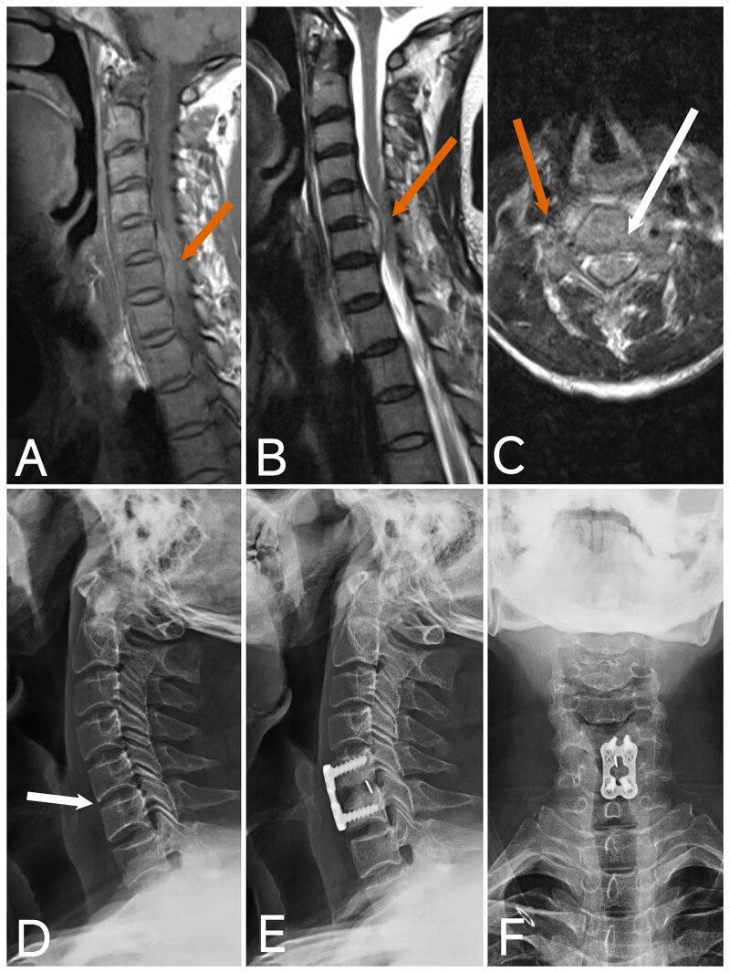
Figure 5.
Image of case 4.
A: T1-weighted imaging (T1WI); B: T2-weighted imaging (T2WI); C: Axial T2-weighted imaging (T2WI) shows an epidural abscess with significant compression of the spinal cord (red arrow). D: X-ray shows no significant bone destruction or loss of intervertebral height (white arrow). E, F: Postoperative X-ray at 4 months demonstrates good positioning of the internal fixation and fusion of the intervertebral bone graft.

