
Fig. 3. Fluorescence localization of wild type and GG4 mutants G202L and G206L of yeast CTR3-GFP.
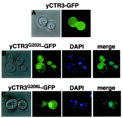
Colonies of yeast transformants of the same constructs used in complementation experiments were spotted directly on glass coverslips for fluorescence microscopy. Bright-field and corresponding fluorescence images are shown. A, plasma membrane localization of wild type yCTR3-GFP fusion protein. GG4 mutants G202L (panel B) and G206L (panel C) are trapped in a perinuclear intracellular compartment as revealed by DAPI staining of nuclear DNA and a ring at the periphery of the cell. Accumulation of GFP-tagged mutant proteins in both regions is indicative of ER localization in yeast. Smaller DAPI-stained points represent mitochondrial DNA.