
Fig. 7. Helical wheel diagram summarizing the results of tryptophan-scanning mutagenesis and complementation experiments.
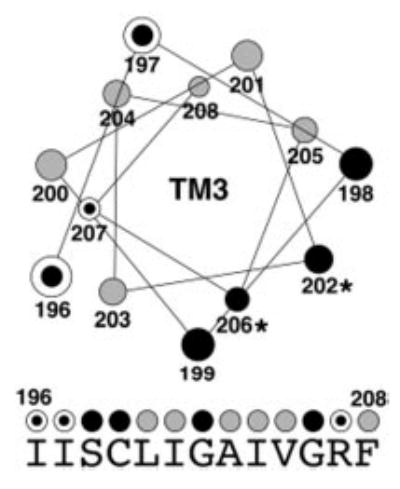
A helical wheel was generated for the middle 13 positions of TM3 (amino acid residues 196–208). Gray circles represent amino acid positions where introduction of a tryptophan residue did not interfere with localization to the plasma membrane and function of the mutant protein in the complementation assay. Three dotted circles identify amino acid positions that caused mislocalization when mutated to tryptophan but were partially functional in the complementation assay. Black circles represent positions where introduction of tryptophan resulted in mislocalization and completely abolished function in complementation experiments. Under the helical wheel, the native sequence of yCTR3 is displayed. The same color-coded circles summarizing the fluorescence localization and complementation results obtained for each tryptophan mutant are placed above each amino acid letter.