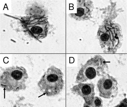
FIG. 3.
Micrographs of J774A.1 cells infected with B. anthracis strains. J774A.1 cells were infected at an MOI of 1 with the indicated strains of B. anthracis. After 8 h, the cells were washed, stained with Giemsa, and photographed using an Olympus IX71 inverted microscope. The panels show representative fields (400× magnification) for B. anthracis Sterne 7702 (A), B. anthracis 9131 (a strain lacking both pXO1 and pXO2) (B), the srtA mutant strain B. anthracis Sterne 7702::pHYZ2 (C), and the srtB mutant strain B. anthracis Sterne 7702::pHYZ3 (D). In panels A and B, heavily stained bacteria are readily apparent. In panels C and D, bacteria (some indicated by arrows) are fewer in number.

