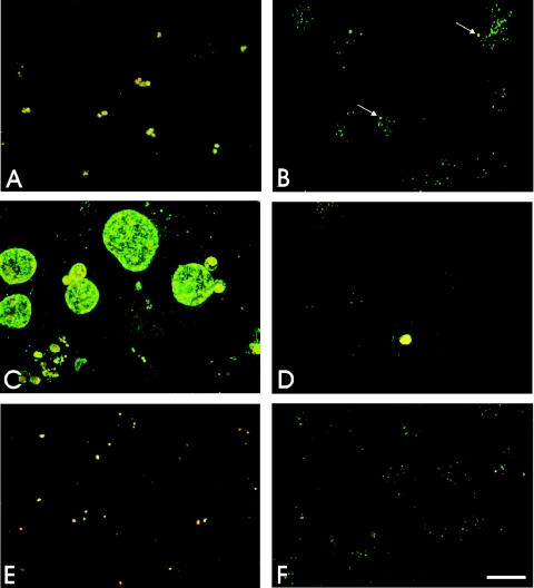
FIG. 3.
(A to D) Colocalization of chlamydial antigens in C. pneumoniae-infected cells. Confocal microscopy of C. pneumoniae inclusions doubly labeled with anti-Hsp60 MAb (red) and anti-C. pneumoniae rabbit antiserum (green) revealed colocalization (yellow) of the antigens to the chlamydial inclusions in HeLa cells at 24 and 72 h postinfection (A and C) and in infected macrophages at 24 and 72 h postinfection (B and D). Only rare inclusions were observed in macrophages (arrows in panel B). (E and F) Colocalization of C. pneumoniae antigens in infected HeLa cells treated with chloramphenicol for 12 h postinfection (F) and in untreated cells (E). Scale bar = 10 μm.

