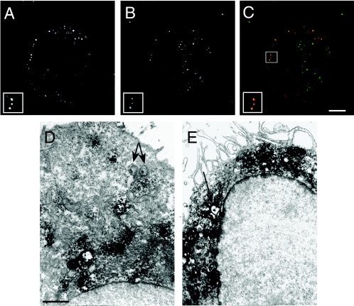
FIG. 5.
Interaction of C. pneumoniae with LAMP-2-positive vesicles. Monocyte-derived macrophages infected with C. pneumoniae were fixed and labeled with anti-LAMP-2 MAb at 2 h postinfection. (A to C) Confocal micrographs revealing colocalization of LAMP-2 with Cell Tracker Green-labeled chlamydiae (A). C. pneumoniae-infected macrophages identifying the organisms (A) and stained for the LAMP-2 marker (B). (C) Composite image showing LAMP-2 (red) and the EBs (green). The insets are higher magnifications of C. pneumoniae colocalizing with LAMP-2-positive vesicles. Scale bar = 10 μm. (D and E) Transmission electron micrographs of C. pneumoniae EBs not colocalizing with LAMP-2-containing vesicles within an infected HeLa cell (D) and colocalizing with LAMP-2-positive vesicles within an infected macrophage (E). The arrows indicate C. pneumoniae EBs. Scale bar = 1 μm.

