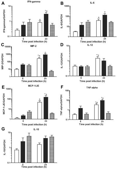
FIG. 4.
Lung mRNA chemokine and cytokine expression in C4 KO mice infected i.t. with serotype 8 pneumococcus: lung mRNA chemokine and cytokine expression as determined by real-time PCR for mice inoculated i.t. with serotype 8 pneumococcus and PBS-treated mice (open bars), IgM-treated mice (solid bars), or D11-treated mice (cross-hatched bars) at 6 and 24 h after infection. (A) IFN-γ. (B) IL-6. (C) MIP-2. (D) IL-12. (E) MCP-1/JE. (F) TNF-α. (G) IL-10. The bars indicate the means of four independent experiments, and the error bars indicate the standard errors of the means. The y axis shows the mRNA concentrations of the mediators normalized to the concentration of glyceraldehyde-3-phosphate dehydrogenase (GAPDH). An asterisk indicates that the P value is <0.05 for a comparison of control and D11-treated mice at 24 h after infection, as determined by an unpaired t test; a plus sign indicates that the P value is <0.05 for a comparison of the mRNA levels at 6 h and 24 h after infection, as determined by an unpaired t test (n = 10 to 12 mice per time).

