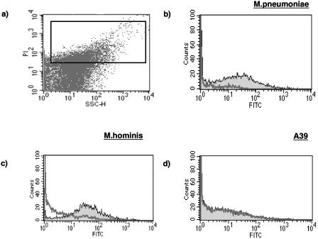
FIG. 1.
Panel a shows a dot plot of a preparation of M. hominis with propidium iodide staining depicted on the y axis and side scatter along the x axis (expressed in arbitrary units). The rectangular acquisition gate is shown. Panels b, c, and d show histograms of three mycoplasma preparations before (thick line) and after (thin line) incubation with MBL. A clear distinction can be seen between organisms that bind MBL (b and c) and the A39 strain (d), which does not. FITC, fluorescein isothiocyanate.

