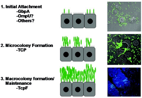
FIG. 8.
Proposed model for V. cholerae intestinal colonization. We propose the following three-step model for colonization: (i) initial attachment (step 1), (ii) microcolony formation (step 2), and (iii) macrocolony formation and maintenance (step 3). Factors that may be involved in each of these steps are listed. Relevant micrographs show GFP-expressing V. cholerae (green) colonizing HT-29 cells (grey) in vitro after 1 and 4 h of incubation; for step 1 the magnification is ×400, and for step 2 the magnification is ×1,000. The micrograph for step 3 shows an infant mouse intestine (blue) colonized by GFP-expressing V. cholerae (green) 16 h after oral inoculation (magnification, ×200).

