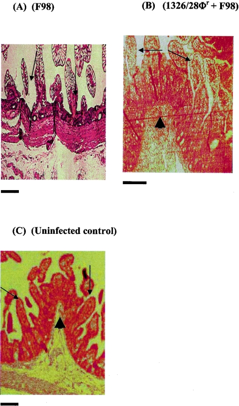
FIG. 6.
Hematoxylin- and eosin-stained ileal sections isolated from F98-inoculated piglets (A), piglets inoculated with 1326/28Φr and then with F98 24 h later (B), and uninfected (control) piglets (C). The arrows indicate long slender villi in panels B and C and stunted villi in panel A. The arrowheads indicate well-formed pilcae in panels B and C. Scale bars = 10 μm.

