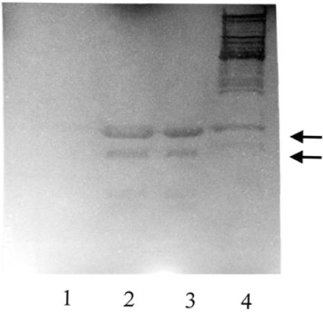
FIG. 3.
Zymographic analysis of bacteriolytic activity of Aaa. The image shows an SDS-PAGE gel of crude cell lysates (10 μl) of noninduced E. coli(pQAaa) (lane 1), IPTG-induced E. coli (pQAaa) (lane 2), six-His-Aaa (1.5 μg) purified from E. coli(pQAaa) (lane 3), and cell surface-associated proteins (10 μl) of S. aureus 4074 (lane 4). The separation gel (12.5%) contained heat-inactivated S. carnosus cells (0.05%) as a substrate for bacteriolytic enzymes. Bacteriolytic activity is visible as a clear zone after incubation in phosphate buffer. The arrows indicate Aaa-associated bacteriolytic activity.

