
Fig. 6. SOCE inactivates at GVBD.
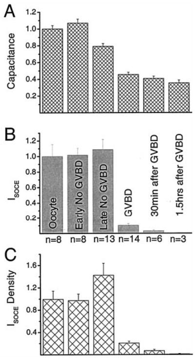
Isoce (A) and cell capacitance (B) were measured at different time points after progesterone addition. C, Isoce density obtained as the current density per unit area (Isoce/capacitance). The data was normalized to values in oocytes because of the variability observed between cells from different donor females. Labeling in B applies to A and C. Early No GVBD refers to cells that have been in progesterone for less than GVBD50, and Late No GVBD refers to cells that have been in progesterone from more than GVBD50 up to 1.3 GVBD50. We measured Isoce in eggs at 0.5, 1, 1.5, 2, 3, 4, and 5 h after GVBD, and no SOCE current could be detected. Data for the 0.5- and 1.5-h time points are shown. The number of cells at each time point is indicated.