
Figure 1.
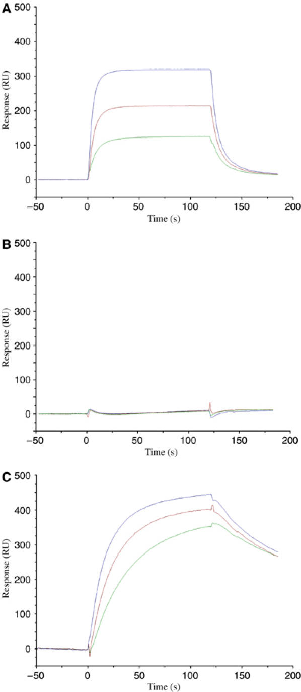
Binding of TCR 3A6 tetramers to MBP/DR2a and K38-27/DR2a complexes. (A) Sensograms showing the binding of 3A6 tetramers at concentrations of 2.3 μM (blue), 1.2 μM (red) and 0.6 μM (green) to HLA-DR2a loaded with MBP 89–101 (VHFFKNIVTPRTP). HLA-DR2a was immobilized on a BIAcore SA chip by injecting 5 μl of biotin-HLA-DR2a (0.1 μM) over the chip. (B) Binding of 3A6 tetramers to HLA-DR2a loaded with an irrelevant peptide. (C) Binding of 3A6 tetramers to HLA-DR2a loaded with K38-27 (WFKLTTTKL), a superagonist peptide (Hemmer et al, 2000).