
Figure 2.
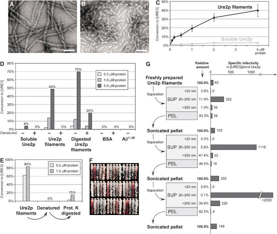
Transformation of in vitro-formed Ure2p filaments into yeast spheroplasts. (A) Electron micrograph of negatively stained Ure2p filaments. Bar, 100 nm. (B) Negatively stained Ure2p filaments after sonication two times for 15 s at 60 W. Bar, 100 nm. (C) Ure2p concentration dependence of conversion to [URE3]. Indicated concentrations of Ure2p filaments and soluble Ure2p were transformed into BY241. Values of [URE3] colonies relative to all Leu+ transformants are the mean of at least three independent experiments (±standard error). (D) Infectivity of proteinase K-digested Ure2p filaments (see Materials and methods) and effect of heat denaturation. Heat denaturation of protein samples was achieved by incubation at 95°C for 5 min. Indicated protein concentrations were transformed into BY241. (E) Recovery of infectivity from heat-denatured Ure2p filaments by proteinase K digestion. Heat denaturation, proteinase K treatment, and transformation were performed as in panel D. (F) Spectrum of [URE3] variants in randomly chosen colonies after transformation of BY241 with Ure2p filaments. (G) Fractionation of freshly prepared Ure2p filaments (see Materials and methods) and specific infectivity following transformation into BY241. The starting concentration of Ure2p filaments was 2 μM; concentrations of each fraction are presented as relative values. Electron micrographs of filament fractions and [URE3] variant spectra of the respective transformants are provided in Supplementary Figure 3.