
Figure 3.
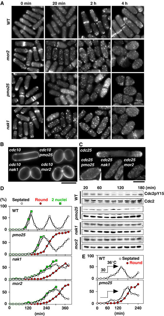
Role of Pmo25 in cell morphogenesis. (A) Cells grown in YES medium at 25°C were transferred to 36°C (time 0), and taken at the indicated times for observation of F-actin localization after being staining with rhodamine-phalloidin. (B, C) Calcofluor (B) and F-actin staining (C) of the double mutants between pmo25 and cdc10 (B) or cdc25 (C). The cells grown at 25°C were shifted to 36°C and incubated for 4 h. (D) Early G2 cells of the mutants collected by elutriation were cultured at 36°C and taken at the indicated times for observation of morphology (left) and for immunoblotting with antibodies specific for Cdc2 phosphorylated on tyrosine-15 (Cdc2pY15) and for PSTAIR (Cdc2; right). (E) Early G2 cells of the mutants collected by elutriation were cultured at 30°C for 1 h and then shifted to 36°C.